-40%
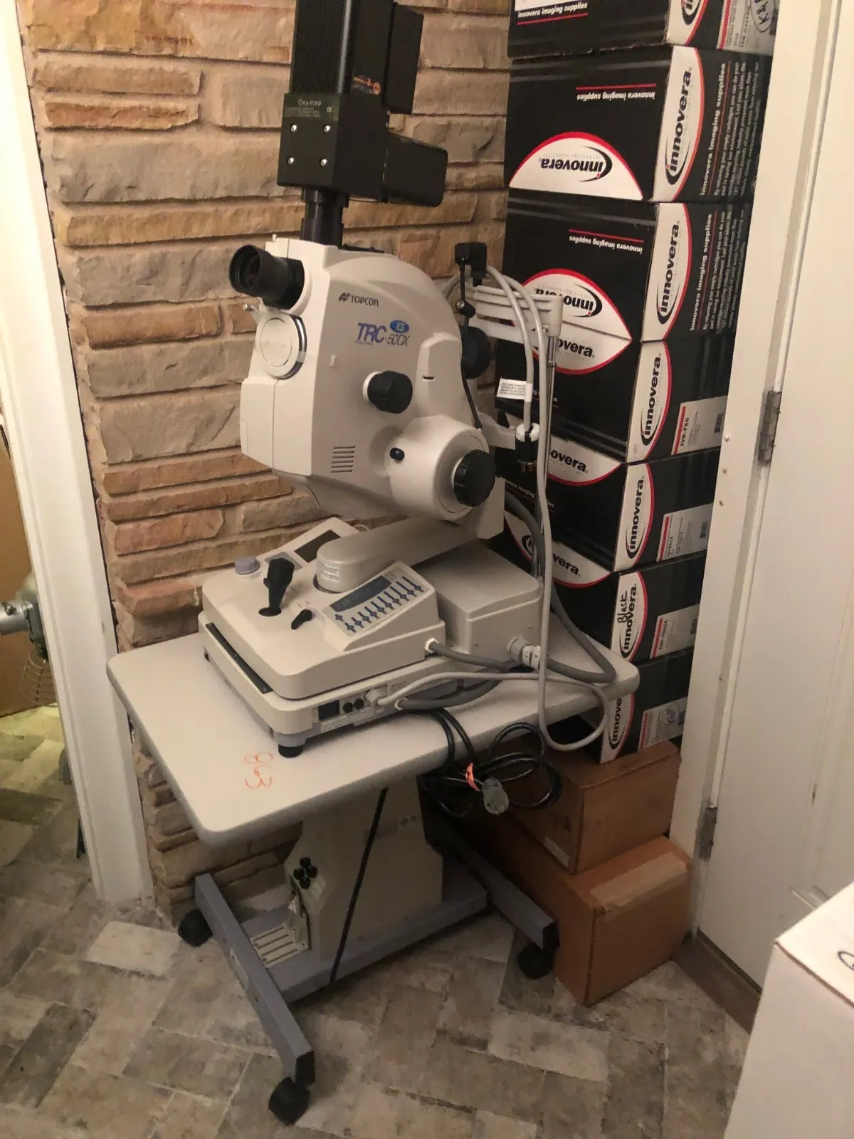
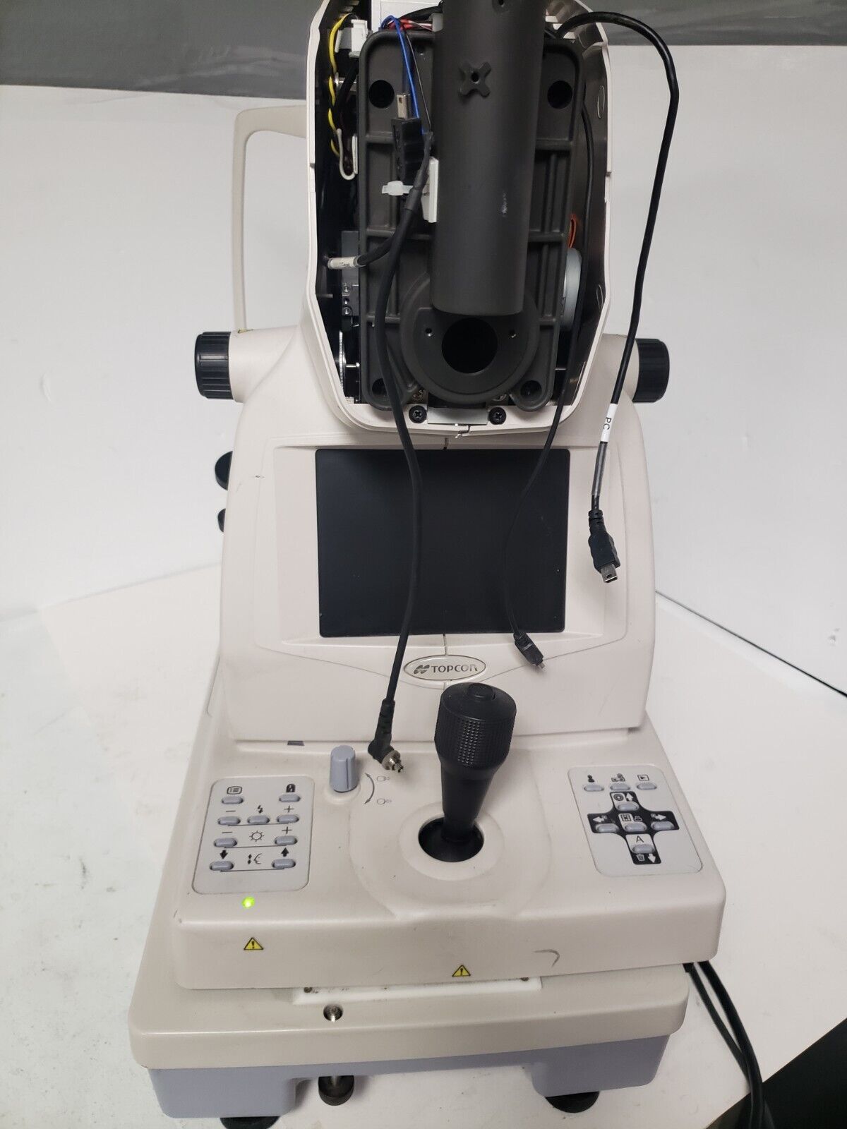
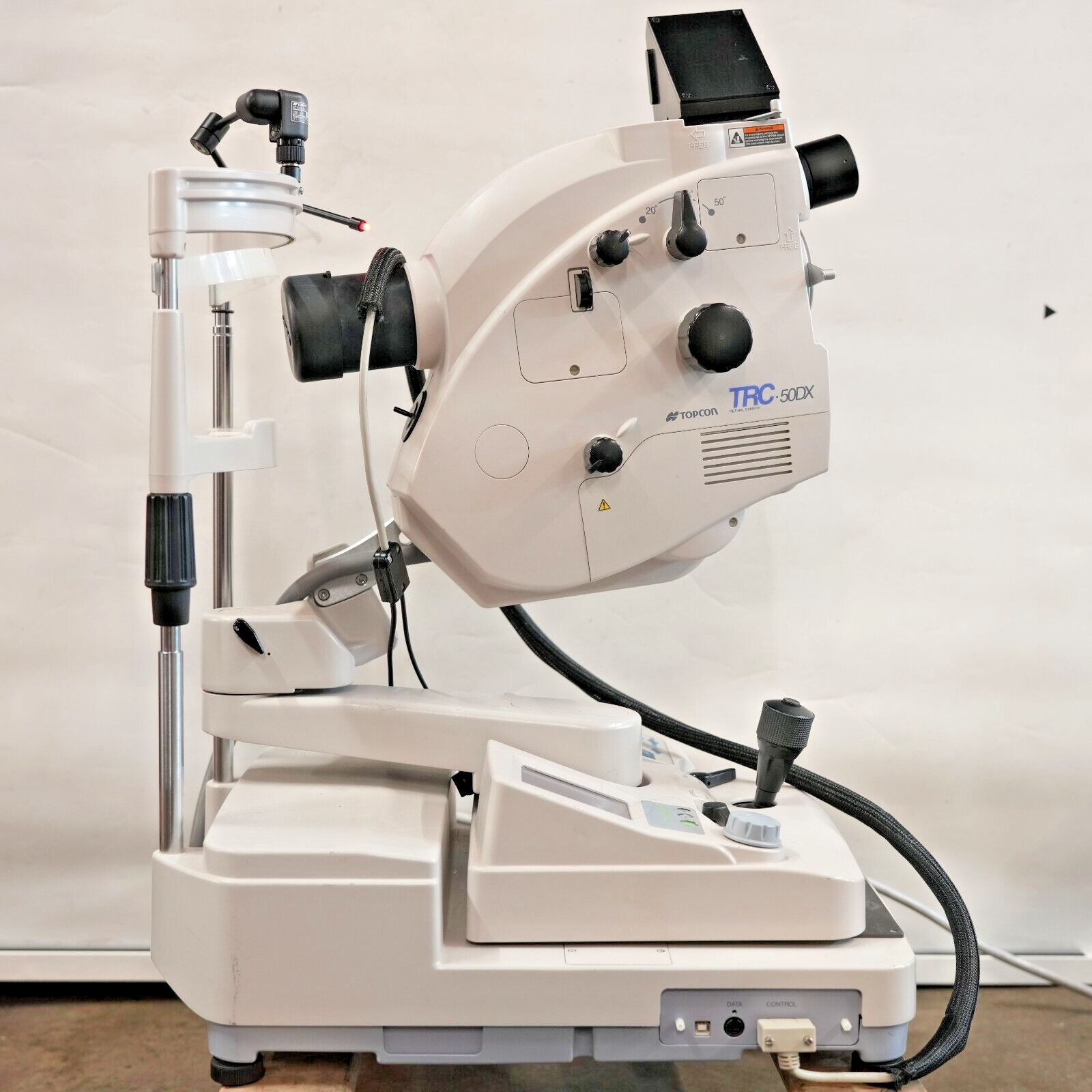
TOP CON - TRC 50DX Mydriatic Retinal Camera with Oxymap Retinal Oximeter
$ 5277.36
- Description
- Size Guide
Description
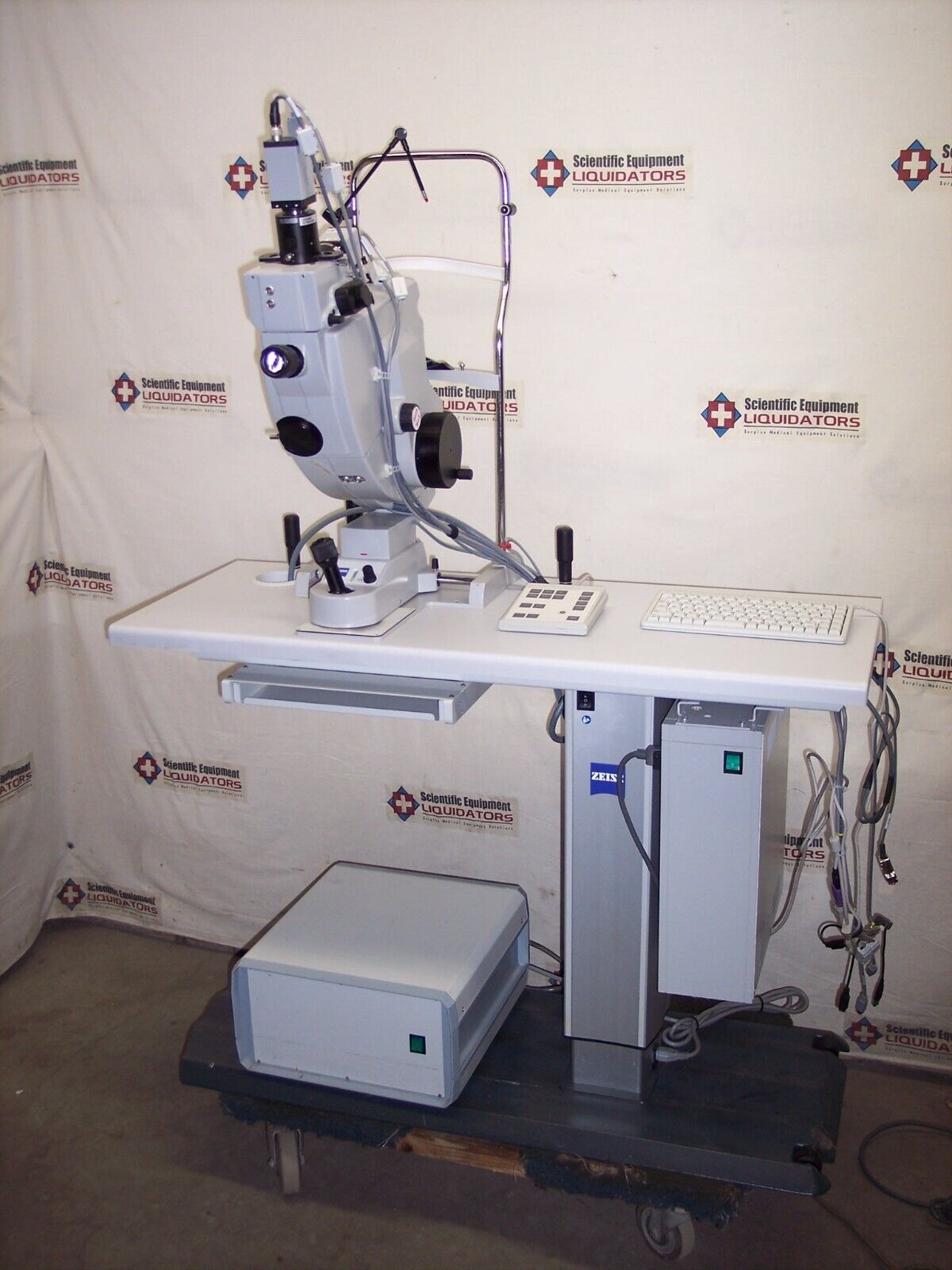
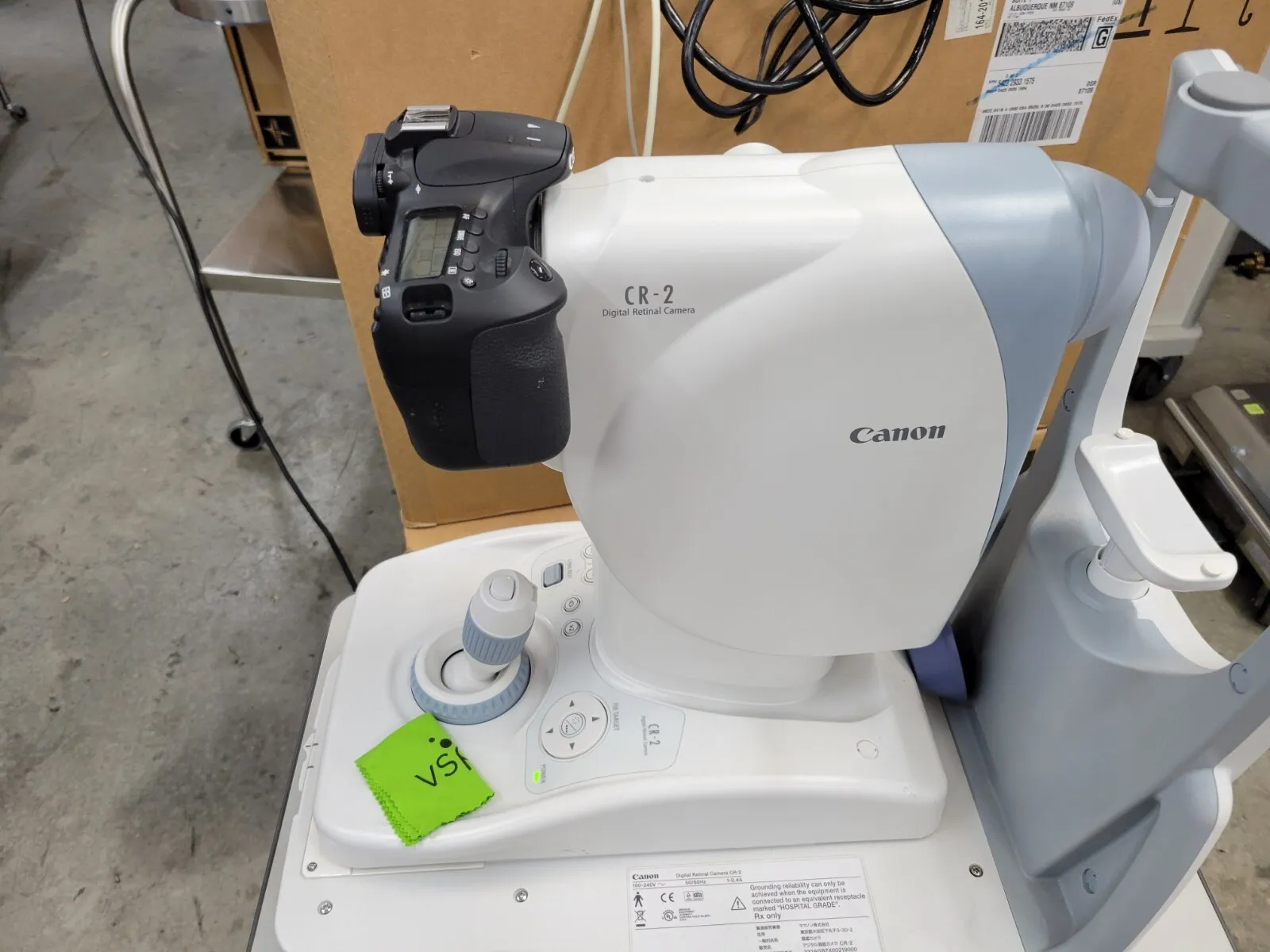
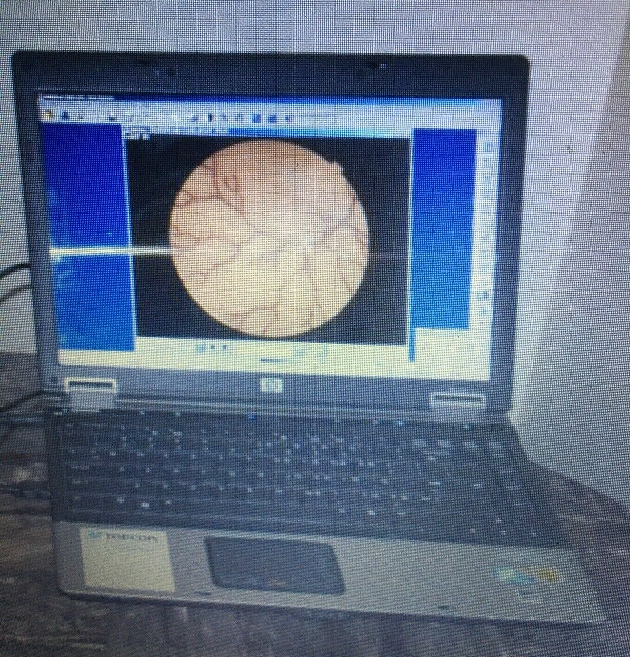
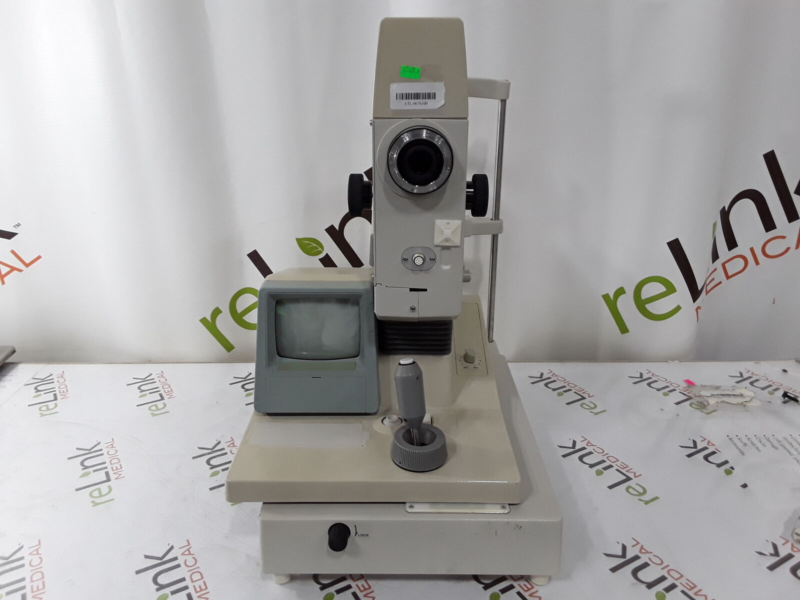
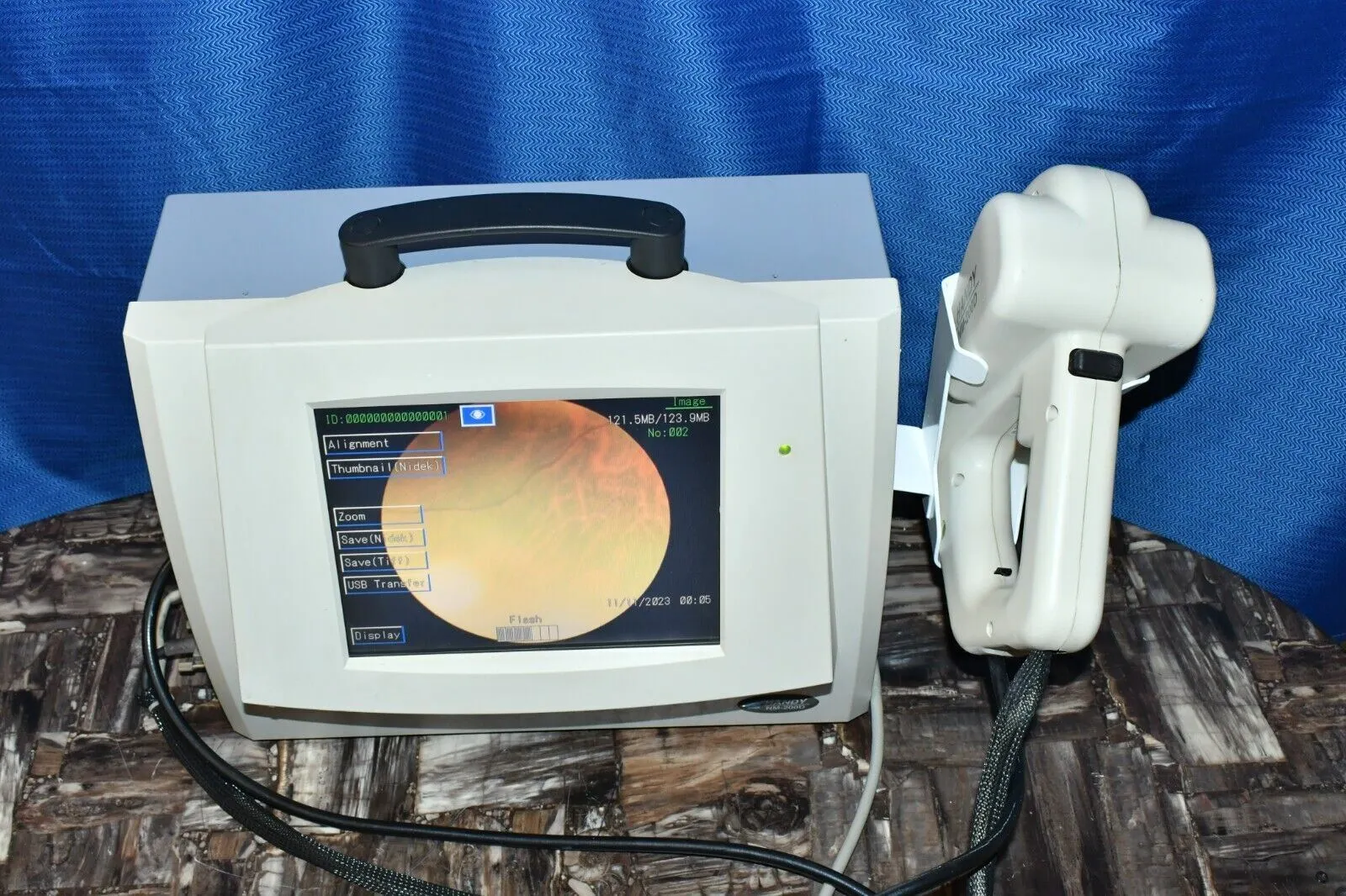
This came from the university of Texas when they finished their study. Includes everything in the listing's pictures. Can ship via freight, and possibly with a regular carrier in parts....local pickup also welcome.The Topcon TRC-50DX improves on the unsurpassed quality of Topcon retinal cameras, incorporating new functions that enhance their versatility and operational ease.
Key Features:
Small pupil mode and aperture adjustment.
Easy-to-use touch-screen control panel.
Comfortable backlit panel for use in darkened environments.
Color fundus, red-free, and fluorescein (ICG and autoflourescence filters are available).
Can support a variety of photo devices, from film to high resolution digital cameras.
50°, 35°, and 20°angles of coverage.
21 levels of flash intensity.
The Oxymap retinal oximeter - visit the following site for more info:
https://www.researchgate.net/publication/224886669_Oximetry_recent_insights_into_retinal_vasopathies_and_glaucoma
The Oxymap retinal oximeter is a spectropho-tometric device based on a fundus camera whichis attached to a beam splitter and a digital cam-era (Figure 1). It simultaneously captures im-ages of the retina at two wavelengths: one issensitive to changes in the percentage of oxy-gen bound to hemoglobin (605 nm) and thesecond is an isobestic wavelength meaning thatlight absorbance is similar for oxygenated anddeoxygenated hemoglobin (586 nm)(4,5,7,11,15,18,19).The fundus view shows colour-coded oxygensaturation values on top of a grayscale retinalimage. In healthy eyes, the arterioles are closeto 90-100% satO2and are coloured in “warm”colours (red to orange) whereas the venules arecloser to 50-60% satO2and are coloured in“cold” colours (green to blue) (18). Special-ized software automatically selects measure-ment points on the oximetry images and cal-culates the optical density (OD, absorbance) ofretinal vessels at both wavelengths, as the log-arithm of the ratio of light intensities inside thevessels versus the outside background. The ra-tio of the two optical densities is approximate-ly linearly related to hemoglobin oxygen satu-ration, since the oxygen saturation is equal tothe percentage of oxygenated hemoglobin with-in total hemoglobin (1,6,7,15-17). Numericalor graphical information of selected vessel seg-ments such as satO2and vessel width are shown(14). Furthermore, it calculates the differencebetween the oxygen delivered to and away fromthe retina (the arteriovenous difference) (11).For this purpose, the method is based on therelationship between light transmittance andoxygen saturation. According to Lambert andBeer, light transmission through a solution di-minishes logarithmically as the concentrationof the solution and the distance through it in-creases (11). Oxygenated and deoxygenatedhemoglobin have different light absorption spec-tra. By analyzing the light absorbance of bloodat these two wavelengths, the oxygenation ofhemoglobin can be estimated. Moreover, Oxy-map is sensitive to changes in oxygen satura-tion. This sensitivity is essential to be of valuefor measuring changes associated with diseaseor treatment
